231 Photos Categorized "Week 4 – Mold ID" (page 1 of 8)
-This fungi was isolated from a purple onion which was in my kitchen fridge. This fungi was labelled 3. -This fungi is a lighter green than it looks…
-This fungi was an air sample isolated from one of the washrooms in my house. This fungi was labelled 7. -This fungi was among the first to appear…
-This fungi was isolated from an onion root, from a white onion that I found in my kitchen. The one I will be describing is the brown fungi….
-This fungi was isolated from dirty rainwater which I found in a puddle outside. The puddle included dirty rainwater, mud, and rocks but I made sure to get…
-This fungi was isolated from a rotting leaf from an unknown large tree in my neighborhood. This rotting leaf was half brown and half green, with some bumpy/rough…
1. Isolated from cheese 2. Appears to be a white, solid, mass colony (or many colonies very closely clustered). Also looks to be flat (not dome shaped). 3….
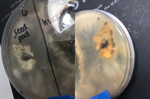
-Isolated from a tree seed pod that was beginning to decay. – It might be two molds because the seed pod is surrounded by a pure white mold…
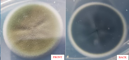
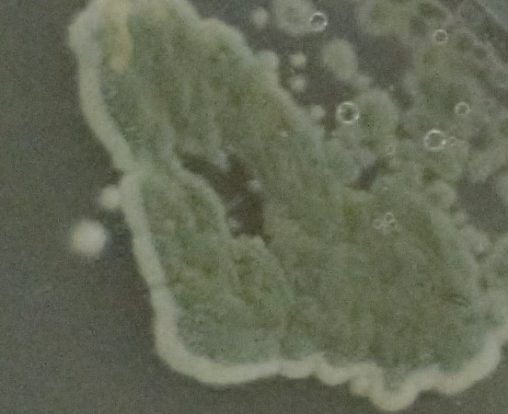
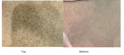
1. Isolated from a soil sample 2. Dull, flat, green, whitish colonies that are clustered together but also spread apart. 3. I’m not sure. This could be trichoderma…
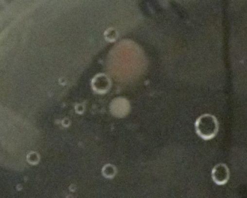
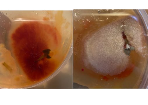
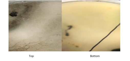
1. Taken from the floor of a standing shower 2. Single, circular colony, pink 3. This is probably not a mold, but the bacterium Serratia marcescens. There is…
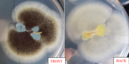
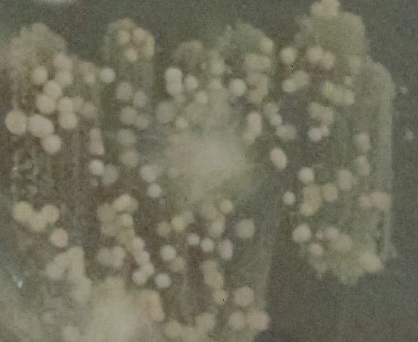
1. Isolated from another cherry tomato 2. Massive green colored colonies clustered together 3. Looks like trichoderma
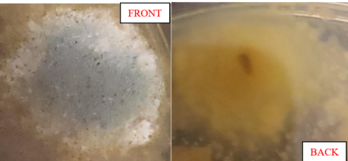
1. Isolated from a cherry tomato 2. Many, far-spread, small, white colonies forming small clusters 3.
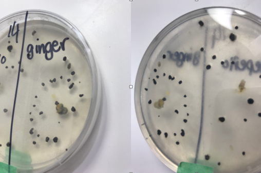
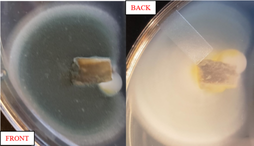
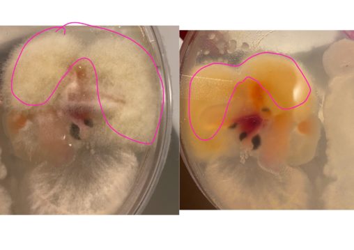
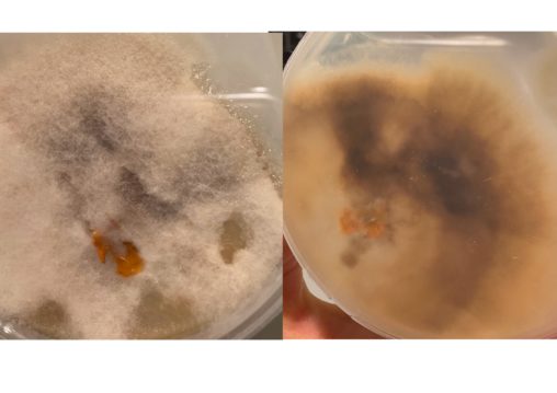
-Mold growing from a sample of ginger. -Transparent mycelium with medium sized, black sclerotia growing in a circle around the ginger sample. -I believe this mold is in…
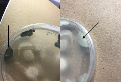
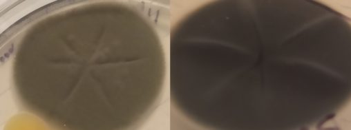
-Also isolated from the air in my apartment. -Grey green on top and white with green edges on the bottom. The mold is growing in five places on…
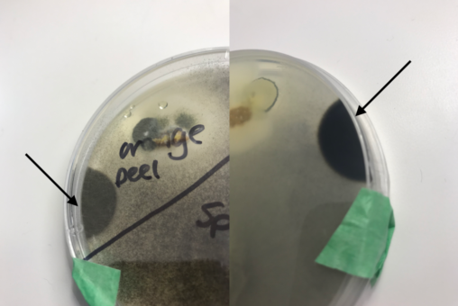
-Grown on a plate with an orange peel and spinach. Although it is closer to the orange peel sample, the spinach really took off and might have spread…
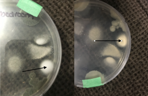
-Mold from an air sample taken from my bedroom/kitchen -The mold is a fuzzy white and pale yellow on the bottom. It was very slow to grow and…

This fungus grew from a swab of rotten squash. It covered the plate in 2 days, and brownish/black sporangiophores lined the lid soon after. Aside from the dotted…
I sourced this fungi from plant tissue from a hosta plant outside in my backyard. THe fungi is fluffy white on top slightly yellow tinged and pure yellow…
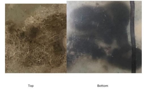
This fungus was growing from a swab taken from a metal fence. It is the same dark brown throughout, growing outward with branch-like mycelia and has a very…
This fungus grew from a kiwi peel and grew for a week before being surrounded by other fungi. It grew its initial colonies in 2 days then expanded…
This was from an air sample on my kitchen window sill behind the sink, left open for an hour. The fungi a bumpy center. It’s a dark green…
This fungus was found from a leaf sample and colonized rapidly. In just 2 days, pigmented colonies were found on the leaf. Each colony was small, and combined…
This was from an air sample on my kitchen window sill behind the sink, left open for an hour. The fungi has cool rings growing from the center….
This fungus grew from a distance away from a withered leaf. It may have come from the leaf, since the leaf moved around a bit when I sampled…
I took a tissue sample from a rotting pumpkin for this fungi. The bottom of this fungi is a dark brown with some of the white/yellowish brownish surrounding…
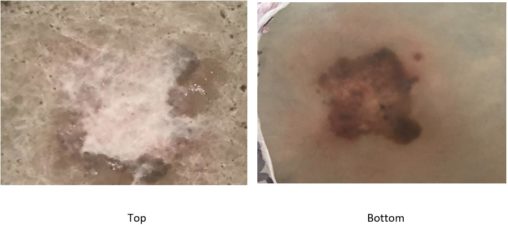
I took a tissue sample from an outer layer of skin from onion. The fungi has long white stalks/ mycelia with black sclerotia on the ends. This fungi…
The material used here is a tissue sample from grass in my front yard. The mold is dark pink on the bottom and white on top. The pink…
This is a sample from moldy inorganic bread on PDA plate. This was one of the fastest growing molds out of all my samples. I think this is…
This is a sample from garden dirt on PDA plate. So many different molds have grown on this plate but I am going to identify the one shown…
This sample is from a moldy cucumber on the PDA plate. on top (not really clear in the picture) the colour is between brown and green /brownish-green and…
This is a sample from kitchen sink on the PDA plate. compared to some of my other samples and my sample from the bathroom sink, this mold grew…