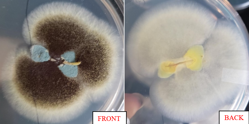
-This fungi was isolated from an onion root, from a white onion that I found in my kitchen. The one I will be describing is the brown fungi. This is labelled slide 13.
-The brown fungi grew in the late second/third week of lab slowly after the blue fungi grew. However, once the brown fungi appeared, the growth rate took off fast and when I would look at the petri-dish at night, it would be a little bigger than what I had seen in the morning. The colour of the fungi is darker brown near the onion root, spreading until it reaches a lighter brown, and then the mycelia is white. However, on the back this fungi is all white with no brown which is interesting. The mycelia in this fungi looks delicate and thin strand-like. The darker brown portion of the fungi almost looks like sclerotia, but when I move the plate around, the tiny speckles don’t move. I remember from the lab that most sclerotia do move.
-This fungi looked exactly like the black mold in the basement slide from lecture. I had three options which included Alternaria alternata, Thielaviopsis basicola, and Stachybotrys chartarum. I decided against Alternaria alternata since the bottom was not dark brown even though it was dark brown at the top fading to light brown edges. It also wasn’t radial but it could have been with more time potentially. I ruled out Stachybotrys chartarum since the bottom was white and not a dark brown, if the top and bottom colours were revered it could have potentially been it. I am IDing this fungi as Thielaviopsis basicola since it looks very similar to the lab slide with brown on top and mostly white on the bottom, but if I look closely there is a darker shade within the white which could be lighter brown on the bottom. There is a darker centre of course with the dark brown at the top. It could be that my fungi had more time to grow than the slide picture in lab which is why there’s more brown than the white mycelia. My fungi does look more delicate but I believe this is the best ID.
