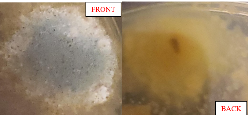
-This fungi was isolated from dirty rainwater which I found in a puddle outside. The puddle included dirty rainwater, mud, and rocks but I made sure to get just the water for my sample. This is labelled slide 14.
-This fungi grew slowly in the first couple of weeks. First all you could see is white but then in the third week you could notice the blue portion in the middle. It’s actually a matt/soft looking textured light-blue but because of my camera quality the zoom makes it look more shiny-ish. The blue portion was slowly spreading from the middle, growing “taking over” the white mycelia. The back, however, looks all yellow. The mycelia in this picture is more blob-like rather than strand-like. This fungi grew in the corner of the plate so it’s slowly coming up the side of the petri-dish. There are also black speckles on this fungi at the top.
– This fungi was difficult to ID because in the lab slides, I didn’t see any that were blue at the top and yellow at the bottom. I had three options for this one which include Penicillium chrysogenum, Aspergillus flavus, or Pestalatiopsis Microspora. I had thought it could have been Aspergillus flavus since the top is a sort-of greenish/blue colour and has some fuzzy white edges. However, it’s not radial, instead it looks like the blue is “taking over” the white so I decided against it. I’m leaning the most towards Penicillium Chrysogenum for the ID because of the distinct blue at the top, which is like the lab slides and one of the slides in lecture Dr. Punja posted. Also, the white edges have almost disappeared since the blue/green is “over-taking it”. However, my fungi is yellow at the bottom. Then I thought it was Pestalatiopsis Microspora since it has a yellow bottom and there are black speckles at the top. However it is not white at the top and is blue. I was stuck between the last two for the ID. The reason I’m going with Penicillium Chrysogenum for this ID is because of the distinct blue/green colour I saw in the slides.