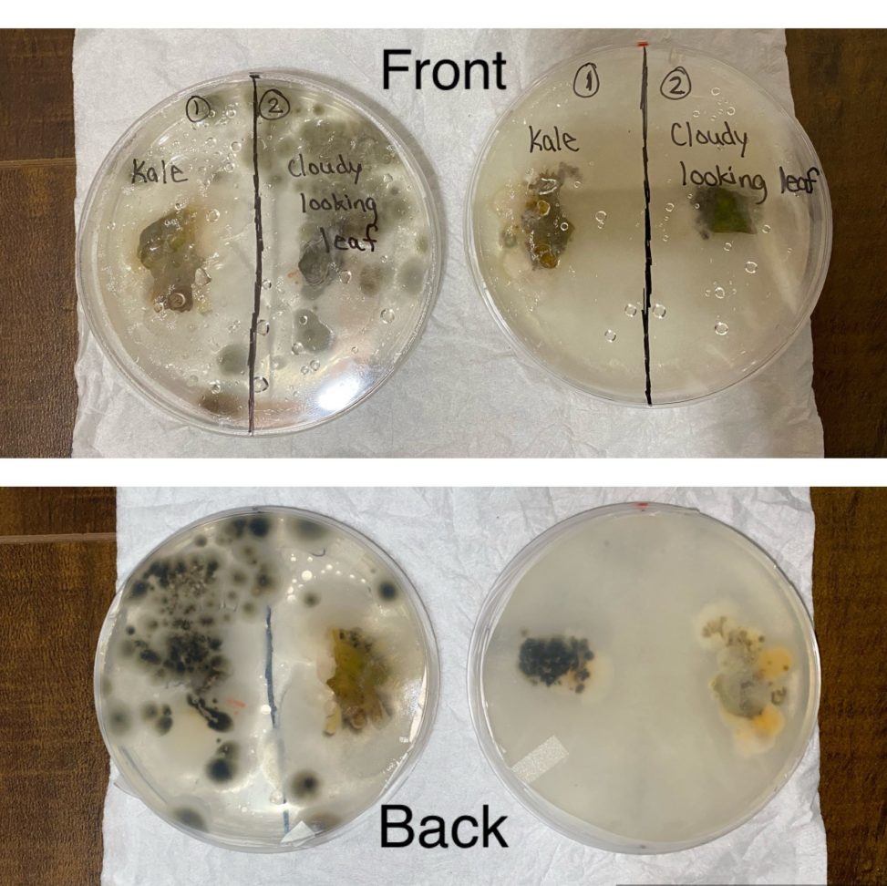
On the plates I have plated samples of wilted yellowish kale (left) and a leaf which had a cloudy covering on it (right side -did not appear healthy compared to other leaves on the plant which had a bright green and waxy appearance). On the right there is a PDA plate and on the left the CMA plate. There are actually a lot of dark greenish colonies present on the CMA plate for the cloudy leaf sample, that they even went on the other side of plate and probably interfered with the growth of the colonies for the kale sample. On the PDA plate the colonies for the cloudy leaf sample are tightly bound to one area. The colonies on the PDA plate are clustered in one area and are smaller in size when compared to colonies on the CMA plate (which are also more scattered on the plate). The kale sample shows about the same amount of growth on both types of media. The only difference I see for the kale sample is that the PDA plate shows more visible yellow fungus growing on it and the CMA plate doesn’t show the yellow fungus growing as clearly as PDA (probably b/c the colonies from the cloudy leaf overtook the space on the plate and didn’t allow the other fungal colonies from the kale to grow as much). I think the CMA media is better for the most part because there are greater amount of colonies grown on that media and they are larger in size than the ones on the the PDA plate.