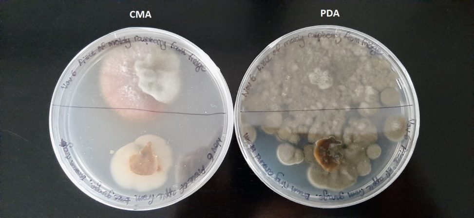
At the top half of both plates is a sample of a moldy raspberry. On the bottom half are samples of apple skin where the skin had turned a leathery brown.
The raspberry sample produced the most fungi growth. On CMA you can see at least 2 white colonies and a single pink colony, although whether the pink colour is the result of the fungus or the raspberry is unclear to me. On PCA, you can see an absolutely horrific amount of greenish-brown mold, which was probably the result of the raspberry sample sliding around my plate when I wrapped it with parafilm ): (It was my first set of plates, ok? Don’t judge). There is so much of it that it had trespassed onto the half with the apple skin sample, making it difficult to tell if the apple skin had any mold growth of its own.
The apple skin on CMA produced more bacteria than mold. I couldn’t see any fungi colonies.
Looking at how much growth the PCA plate houses, I think it’s safe to say that it’s a better media type for the fungi found on the raspberry.