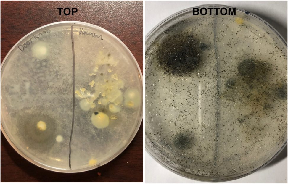
The source material that I decided to isolate this fungi from came from a swab on a doorknob handle and a little bit of vacuum dust. These photos were taken from the CMA plate. This fungi was fast growing, where it covered the whole petri dish in less than a week. There is white mycelium with black spots growing on the top of the strands that are filling the whole petri plate. This fungi is a bit hard to see in the photo due to the condensation but from what I can see, I believe this mold is Rhizopus stolonifer. This is because the fungi I isolated and R. stolonifer both have the same macromorphology, which is white mycelium containing black spots at the tip of the strands. From what I can see, there is no differences between the fungi I isolated and R. stolonifer.
