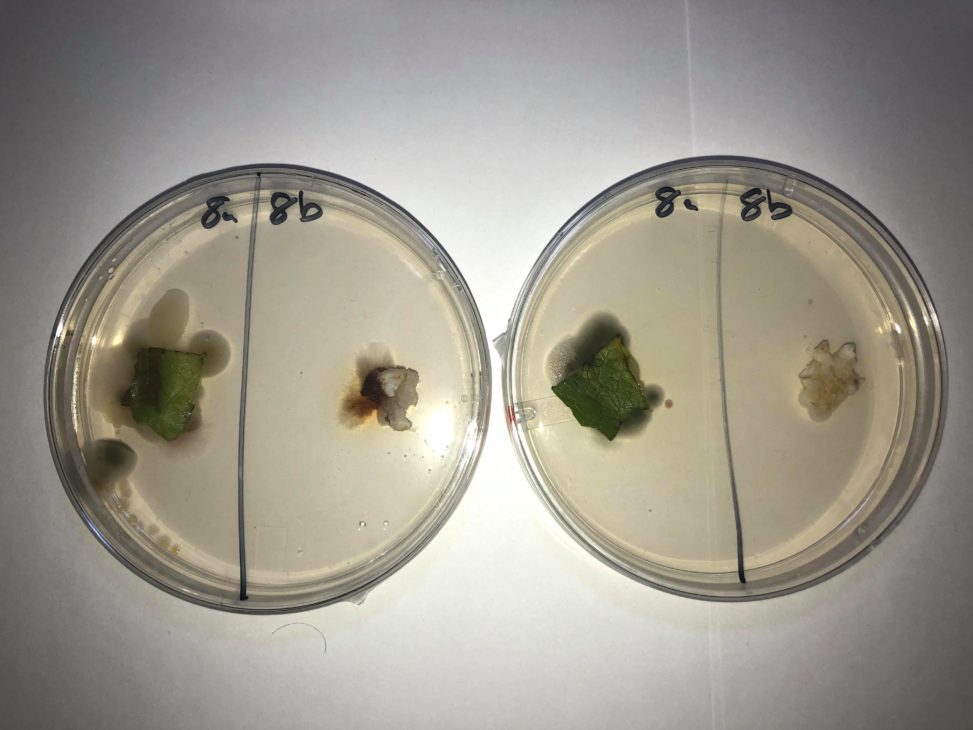
These plates contain a romaine lettuce sample (8a) and a rice cracker sample (8b). The CMA plate is on the left, the PDA plate on the right. There seems to be more growth on the CMA plate, and more variety; there are multiple areas of different size fungal growth. The fungi on the rice cracker is a bit darker red on CMA than PDA so perhaps it is closer to its maximum growth and is producing secondary metabolites. I believe CMA is a better medium for these samples as their growth seems to be better.
