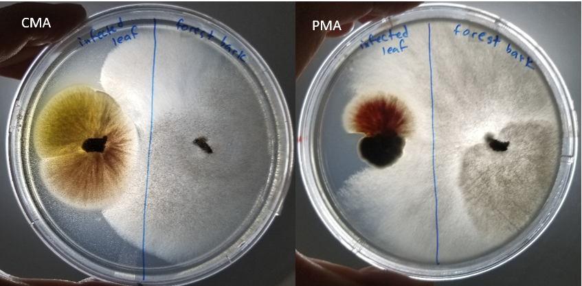
The photo was taken against the light to see the beautiful mycelia of the fungus, and the photos show the front side of the plate. Samples consist of the infected leaf on the right of the plate and forest bark on the left of the plate. CMA plate is on the left while the PMA plate is on the right. The PMA media allowed the forest bark to grow rapidly inhibiting the growth of fungus from the infected leaf. In the CMA plate, the infected leaf was able to grow to a greater extent compared to the PMA. Based on the growth, the PMA is a better medium to isolate fungus from forest bark. For the fungus of the infected leaf, the media choice is inconclusive since the growth in PMA was hindered by the white fungus from the forest bark sample.