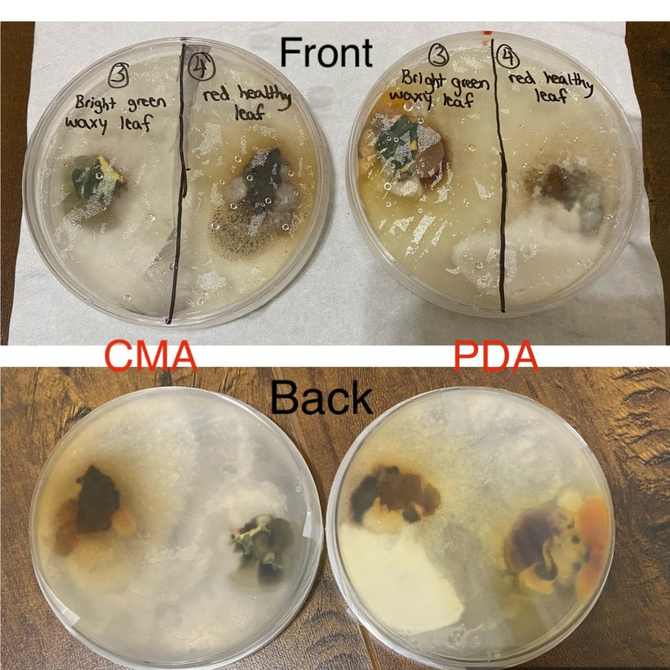
On the left side there is a sample of a healthy bright green waxy leaf taken from a plant which also had unhealthy cloudy looking leaves. On the right side there is a sample of a healthy red leaf taken from a tree outside my house. Both the plates show about the same amount of growth for both the leaves. However, there is slightly more more growth on the PDA plate for the waxy green leaf sample. Also for the green leaf sample more types of fungus is visible on the PDA plate like orange, yellow and small black colonies. Whereas, on the CMA plate for the green leaf sample not much fungal growth is visible except a dark greenish blob which has not spread that much either. But for the both the plates there is white fungus growing from the green leaf sample. For the red healthy leaf, both plates show the same type of fungal colonies(white, brown, peachy yellow) from which the the white cloudy fungus is growing from the read leaf onto the other side of the plate. There is also about the same amount of growth of fungus for most of the colonies except the white one (more on the PDA plate). Overall, I think the PDA plate is better for both leaf samples because for the red leaf there is more growth of the white fungus covering pretty much the other half of the plate. And for the bight green leaf, the PDA plate shows more variety of different types of fungal colonies (whereas CMA shows only black and white).