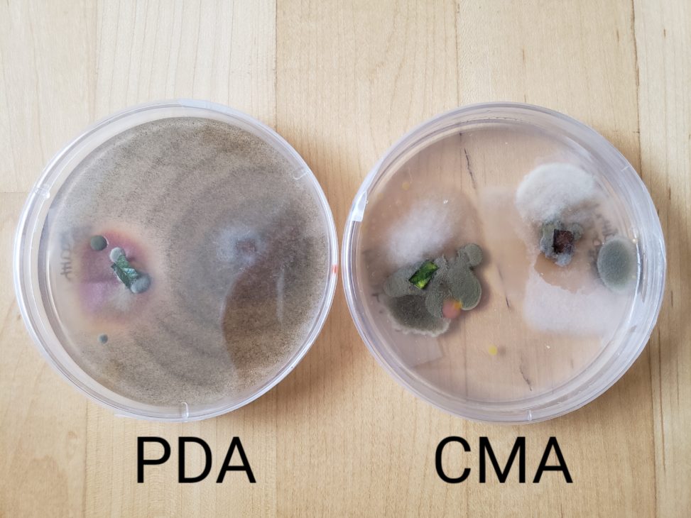
Two leaf samples from two different strawberry plants (one healthy, one unhealthy) were plated on the petri dishes. The leaf tissue on the left side of the plates is from a relatively healthy strawberry plant with no red leaves, and the samples plated on the right side of the PDA and CMA plates are from an unhealthy strawberry plant with red leaves. In the PDA plate, you can see that there is one fungi from the unhealthy plant that is quickly taking over the plate, but the same fungi can’t be seen on the CMA plate. On the healthy leaf tissue side in the PDA plate, you can see around three different fungi, but they have a slower growth rate compared the fungi on the other sample. There is also a red fungi that is seen in the PDA plate but not the CMA plate. In the CMA plate, it looks like there are similar fungi from both the healthy and unhealthy leaf, and they are growing at around the same rate. I think the PDA media was better because it allowed a more diverse set of fungi to grow on the plate compared to the CMA media. It also allowed the brown fungi to grow really quickly. The CMA media looked like it was also contaminated with bacteria which isn’t ideal if we want to solely isolate fungi.
