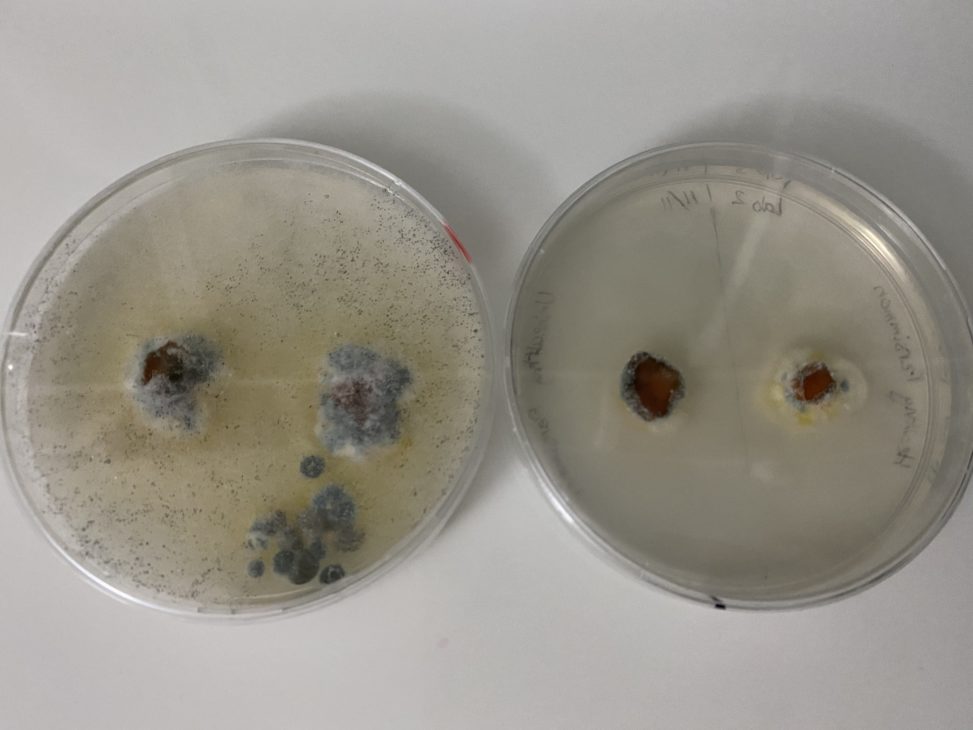
A sample of a persimmon with a brown spot was plated on the left section of both plates. The right section of both plates contains a sample of a healthy persimmon. PDA on left side, CMA on right side. The left PDA plate has very visible signs of black fuzzy mycelia and hyphae all over the plate on both samples, while the right CMA plate lacks this. The CMA plate has small amounts of black fuzzy mold around the edges of the unhealthy persimmon sample, while the healthy persimmon contains white fuzz. The PDA media appears to be better, because a large amount of visible mycelia is growing all over the plate, while the CMA plate only has localized mold growing on just the edges of the persimmon.
